Fig. 2.
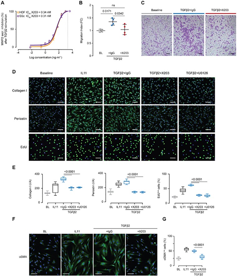
IL11 antibodies block TGFβ2-driven activation of skin fibroblasts from SSc patients and controls
(A) Dose–response curve and X203 IC50 value in inhibiting MMP2 secretion by HDFs and SSc dermal fibroblasts stimulated with TGFβ2. (X203 serial 4-fold dilution from 4 µg/ml to 61 pg/ml; line at x = 2 µg/ml; total n = 2 per group.) (B) Transwell migration indices of HDFs following 24 h stimulation with TGFβ2 (5 ng/ml) and in the presence of IgG or X203 (2 μg/ml). Data: n = 4, mean with s.d. Statistics: one-way ANOVA, Tukey’s multiple testing correction. (C) Migration assay representative images; scale bar, 150 µm. (D) Representative images of collagen I, periostin and EdU stainings of HDFs, following a 24 h stimulation with IL11 (5 ng/ml), or a combination of TGFβ2 (5 ng/ml) and IgG (2 μg/ml), IL11 neutralizing antibody (X203, 2 μg/ml) or ERK inhibitor (U0126, 10 μM); scale bar, 100 µm. (E) Quantification of signal intensity/area (collagen I, periostin) and percentage of EdU-staining positive cells relative to experiment in (D). Statistics: one-way ANOVA, Tukey’s multiple testing correction. (F and G) Representative images and quantification of percentage of αSMA-staining positive cells following a 24 h stimulation of SSc fibroblasts with the indicated factors. Data: whiskers from min. to max. value, line at median. Statistics: one-way ANOVA, Tukey’s multiple testing correction. αSMA: α-smooth muscle actin; EdU: 5-ethynyl-2′-deoxyuridine; ERK: extracellular signal-regulated kinase; HDF: human dermal fibroblast.

