Fig. 3.
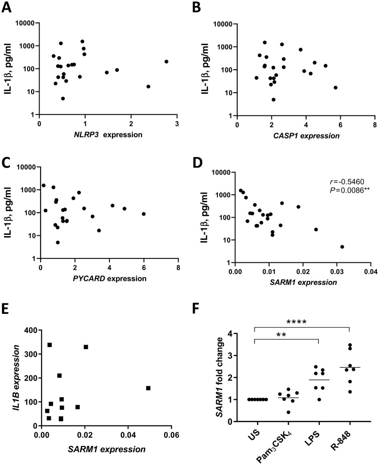
SARM expression negatively correlates with IL-1β secretion following TLR1/2 stimulation in RA monocytes
TLR1/2-induced IL-1β secretion was measured following 24 hours of stimulation with 100 ng/ml Pam3CSK4 and correlated with the basal expression of (A) NLRP3 (n = 22), (B) CASP1 (n = 22), (C) PYCARD (n = 22) and (D) SARM1 (n = 22) in monocytes from RA patients. The significance was analysed using a two-tailed Spearman test. (E) Monocyte IL1B expression following a 3-hour stimulation with 100 ng/ml Pam3CSK4 was compared with basal SARM1 expression (prior to stimulation) in matched patient samples (n = 12). (F) Monocytes from healthy volunteers (n = 7) were stimulated for 6 hours with 100 ng/ml Pam3CSK4, 10 ng/ml LPS or 2 µg/ml R-848. SARM1 expression was expressed as the fold change from US cells and shown as the mean ± SEM. A one-way ANOVA using Dunnett’s multiple comparisons test was used to test significance compared with US cells (**P = 0.007, ****P < 0.0001). LPS: lipopolysaccharide; SARM: sterile-α and armadillo motif containing protein; TLR: Toll-like receptor; US: unstimulated.

