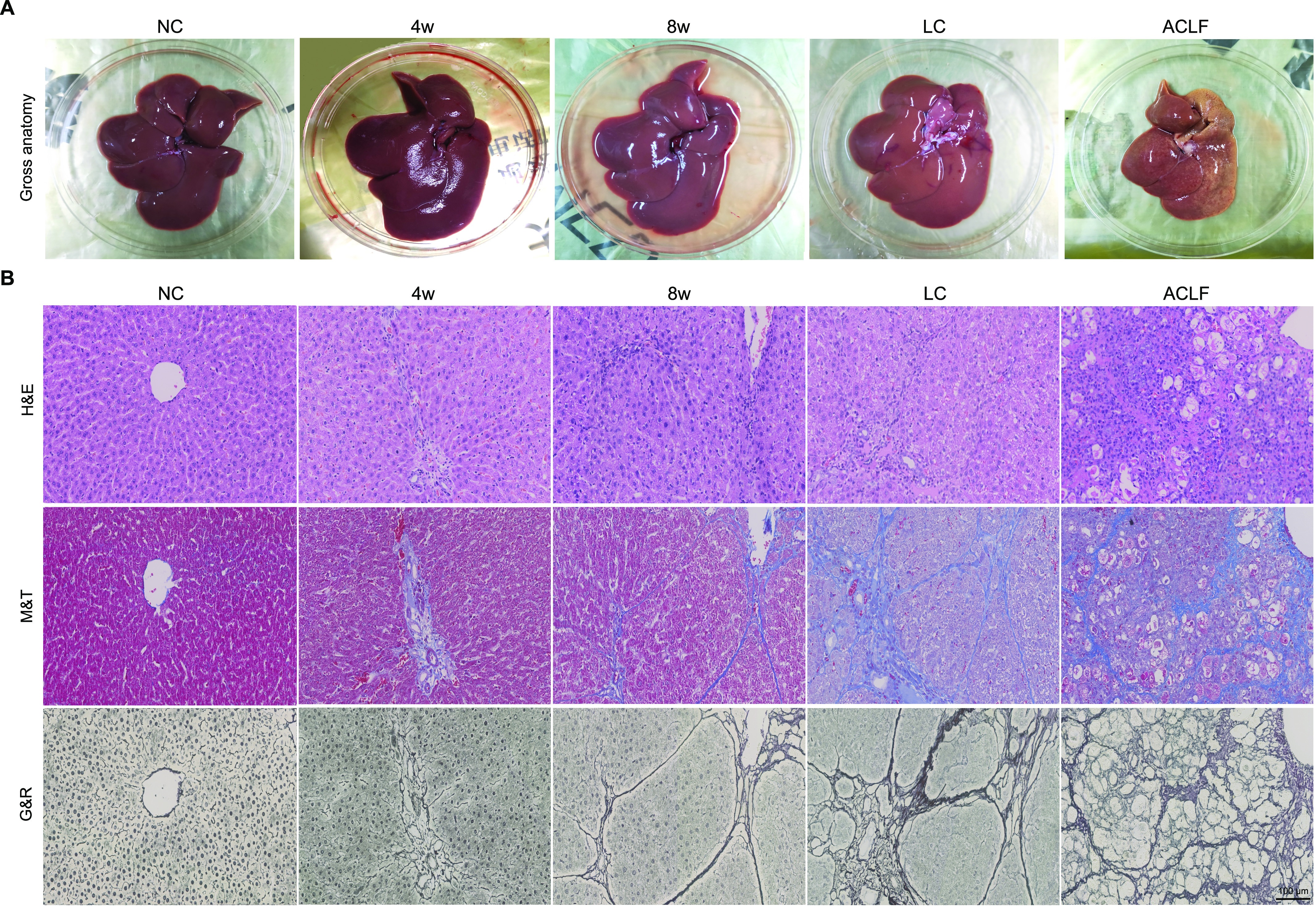
Figure 2.

Morphological and histopathological assessment of liver samples. (A) Liver morphological changes in NC, LC, and ACLF rats. (B) Representative images of the pathological staining (H&E, M&T, and G&R) of liver sections collected from rats at different disease stages (bar = 100 μm). ACLF, acute-on-chronic liver failure; G&R Gomori’s reticulin; H&E, hematoxylin-eosin; LC, liver cirrhosis; M&T, Masson’s trichrome; NC, normal control.
