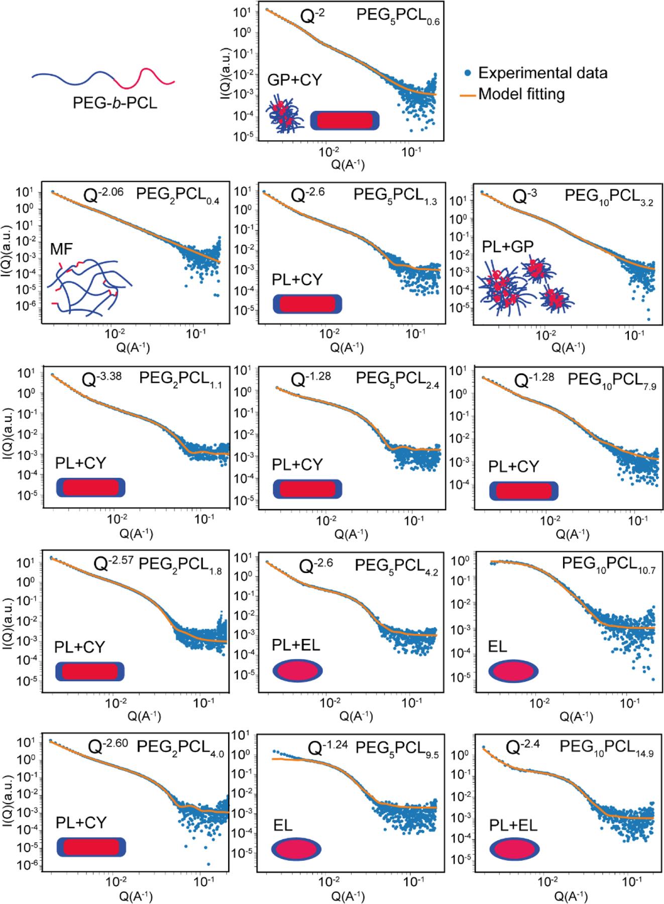
Figure 6:

Synchrotron small-angle X-ray scattering profiles and model fitting for PEG-b-PCL block copolymers in water. Illustrations indicate the proposed structure of the aggregates, wherein the PCL cores are depicted as red, and the PEG coronas are shown in blue. The models which have been applied for fitting are denoted as follows: MF = mass fractal (random-walk polymers loosely clustered), PL = power-law scattering from secondary polymer aggregates, CY = cylindrical micelles, GP = Guinier-Porod (collapsed polymer chain), EL = ellipsoidal micelles.
