
Figure 5:
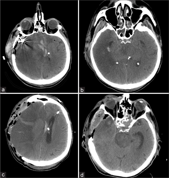
(a and b) computed tomography (CT) of the head on the 3rd day after the surgery. Hemispheric ischemic impairments on the right, expressed edema and dislocation of the brain. (c and d) CT of the head after decompressive trepanation. Expressed brain prolapse into the trepanation defect. Cisterna ambiens is visualized.