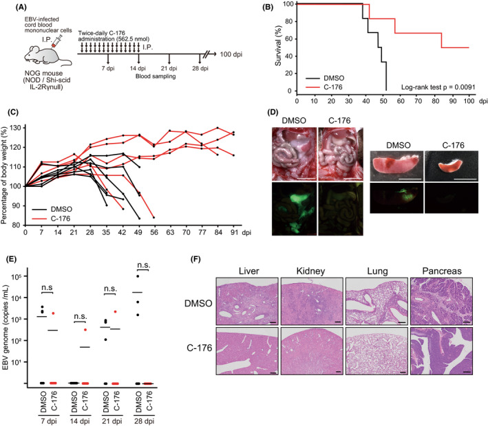
FIGURE 1.

STING inhibitor suppresses EBV tumor formation in the EBV‐LPD mouse model. A, Schematic of the mouse model. A NOG mouse was inoculated ip with EBV‐infected human cord blood mononuclear cells and then administered DMSO or C‐176 (562.5 nmol) ip, twice daily for 2 wk (n = 6 mice per group). Blood sampling was carried out to identify genomic EBV DNA at the indicated time points. B, Kaplan‐Meier survival curves. C, Bodyweight changes of mice are shown. D, The ip tumor and spleen of DMSO‐treated and C‐176‐treated mice. The EBV‐encoded EGFP signal was detected under excitation light. Scale bars, 5 mm. E, The EBV genome (copies/mL) of each group is indicated. EBV DNA in blood was detected by real‐time PCR. n.s., no significant difference. F, H&E staining of DMSO‐treated and C‐176‐treated mouse organs. Scale bars, 200 μm
