FIGURE 5.

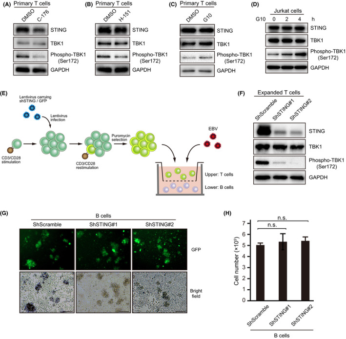
STING inhibitors suppressed STING signaling in T cells. Immunoblot analysis showing STING, TBK1, phospho‐TBK1 (Ser172), and GAPDH. A, B, Primary T cells were isolated from PBMCs and reacted with 0.5 µM C‐176 or 0.5 µM H‐151 for 24 h. C, Primary T cells were exposed to DMSO or 50 µM G10 for 2 h. D, Jurkat cells were treated with DMSO or 50 µM G10 and harvested at the indicated times. E, Human CBMC‐derived B cells were co‐cultured with EBV in vitro. On day 1 after infection, expanded T cells carrying shScramble or shSTING (#1 and #2) were seeded into the transwell chamber. F, Immunoblots for endogenous STING in shSTING‐transduced T cells. G, Fluorescence image of B cells co‐cultured with T cells. Scale bars, 200 μm. H, B cells were quantified at 7 d after infection by counting trypan blue‐stained cells
