
FIGURE 2.
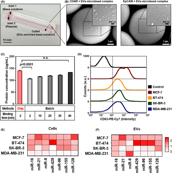
Enrichment of EV‐miRNA by microfluidic chip. A, Image of HOMM microfluidic chip. B, Scanning electron microscope (SEM) image of MDA‐MB‐231 tumor‐derived EV (TDE) after hydrodynamic separation using the microfluidic chip. The magnified SEM image shows TDE bound to the surface of 7‐μm beads coated with EpCAM and CD49f‐specific antibodies. C, Time‐dependent TDE isolation efficiency using the microfluidic chip compared to the batch method of IP. D, Flow cytometry of EpCAM and CD49f‐specific TDE after immunostaining with CD63 fluorescently labeled antibodies. E, Heat map of miRNA expressions for breast cancer cell lines. F, The specific EV enriched by the microfluidic chip