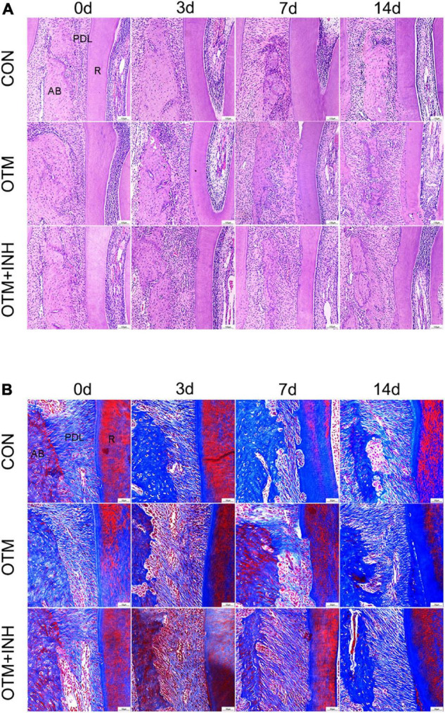
FIGURE 3.

Histological changes of PDL on the tension side of the distobuccal roots. HE staining (A) and Masson’s trichrome staining (B) of PDL. Periodontal fibers were stretched by applying orthodontic force in the OTM group, while remodeling was delayed by the inhibitor of Piezo1 in the OTM + INH group. Scale bar for (A) = 100 μm. Scale bar for (B) = 50 μm. AB, alveolar bone; R, root.
