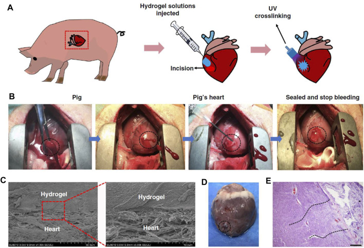
FIGURE 3.
Hemostatic properties of the matrix gel in a pig cardiac puncture injury model. (A) Schematic diagram of the surgical procedure. (B) Gross view of the rapid hemostasis and sealing following cardiac puncture injury. (C) Scanning electron micrographs of the interface between the pig heart puncture wound and the hydrogel. Scale bar: 50 μm (left plates); 10 μm (right plates, enlarged). (D) Images of a heart autopsy following killing after two 2 weeks of postoperative recovery, the hydrogel still adhering to the wound. (E) Tissue staining images of the interface between pig heart cardiac tissue and the matrix gel, after 2 weeks of postoperative recovery. Scale bar: 200 μm (n = 4). Reproduced from (Hong et al., 2019) with permission from Copyright 2019 Springer.

