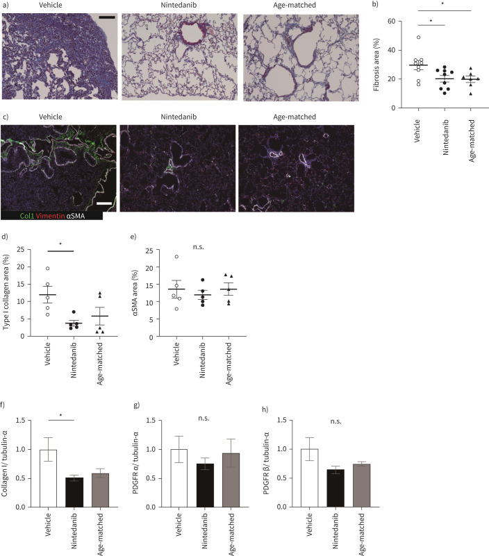
FIGURE 2.
Nintedanib attenuates lung fibrosis. a) Masson's trichrome staining for vehicle, nintedanib-administrated or age-matched lung specimen. b) All images for the section from left lung and accessory lobe are captured at a magnification of 200×, and blue fibrotic areas for the total lung areas are calculated by Fiji. Data are presented as mean±SE of nine mice for each group. c) Immunohistochemical staining for Type I collagen (Col1, green), vimentin (red) and αSMA (white). Scale bars=100 μm (white and black). d) and e) Type I collagen (d) and αSMA (e) expressing areas in left lung and accessory lobe are calculated by Fiji. The calculation of stained images is done in the same way as in (b). Data are presented as mean±SE of five mice for each group. f–h) Type I collagen (f), PDGFRα (g) and -Rβ (h) in the whole lung tissues are assessed by Western blotting. Data are presented as mean±SE of four mice for each group. *p<0.05 compared with vehicle group. n.s.: nonsignificant.

