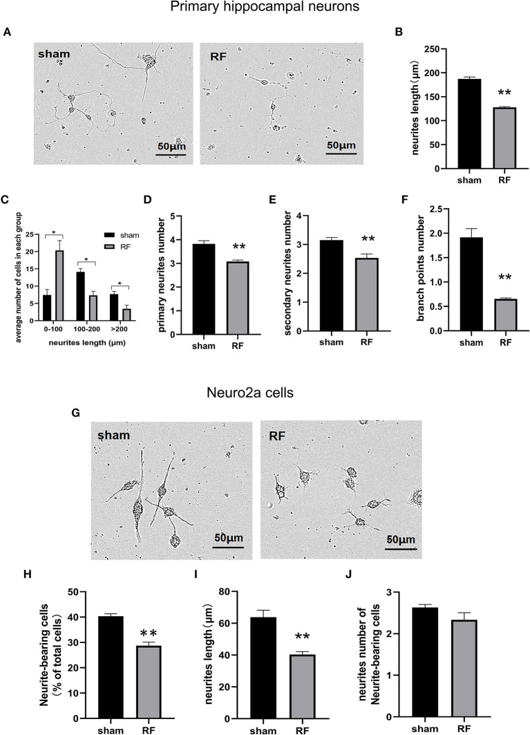
Figure 2.
Effects of RF-EMR exposure on neurite outgrowth in neuronal cells. Primary hippocampal neurons (A–F) and Neuro2a cells (G–J) were exposed to 1,800 MHz RF-EMR at 4 W/kg for 48 h. Morphological parameters were identified according to the description in the methods. Representative images of primary hippocampal neurons (A) and Neuro2a cells (G) were taken under a (20 ×) Leica microscope, and pictures were analyzed with ImageJ software. (B) shows the total neurite length per primary hippocampal neuron, (C) shows the number of cells in the groups with a total neurite length per primary hippocampal neuron larger than 100 μm, (D,E) shows the primary and secondary neurite numbers per primary hippocampal neuron, (F) shows the branch point number per primary hippocampal neuron, (H) shows the neurite-bearing cell percentage in Neuro2a cells, (I) shows the total neurite length per Neuro2a cell, and (J) shows neurite number of neurite-bearing Neuro2a cells after RF-EMR exposure. *P < 0.05, **P < 0.01, Student's t-test. Scale bar: 50 μm.

