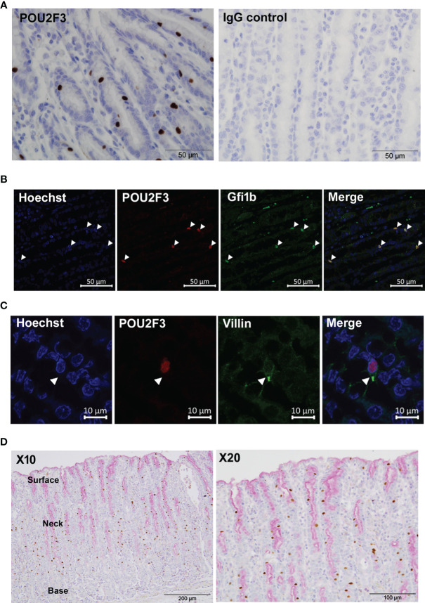
Figure 1.
Identification of tuft cells in ovine abomasal epithelium by immunohistochemistry. (A) Detection of tuft cells using antibody to human POU2F3 (brown) in ovine abomasal epithelium sections at day 21 post-infection with Teladorsagia circumcinta (left hand panel). Surrounding epithelial cells are counterstained with hematoxylin blue. IgG isotype control antibody showed no labelling (right hand panel). (B) Co-localization of antibodies to POU2F3 and GFI1b in ovine tuft cell nucleus. Arrowheads indicate nuclear localization of each antibody individually and in the merged image. Nuclei were stained with Hoechst. (C) Detection of apical tuft on POU2F3+ cells (red) with antibody to villin (green). Arrowhead indicates tuft-like structure on double-labelled cell. Nuclei were stained with Hoechst. (D) POU2F3+ cells are distinct from mucin-secreting cells. Co-labelling with POU2F3 antibody (brown) and mucin stain Periodic Acid-Schiff (pink), which localizes to the surface mucus layer and to surface and neck mucous cells, predominantly at the apical (upper) edge of the abomasal epithelium. Areas of surface and neck mucous cells and the base of the gastric glands are annotated.

