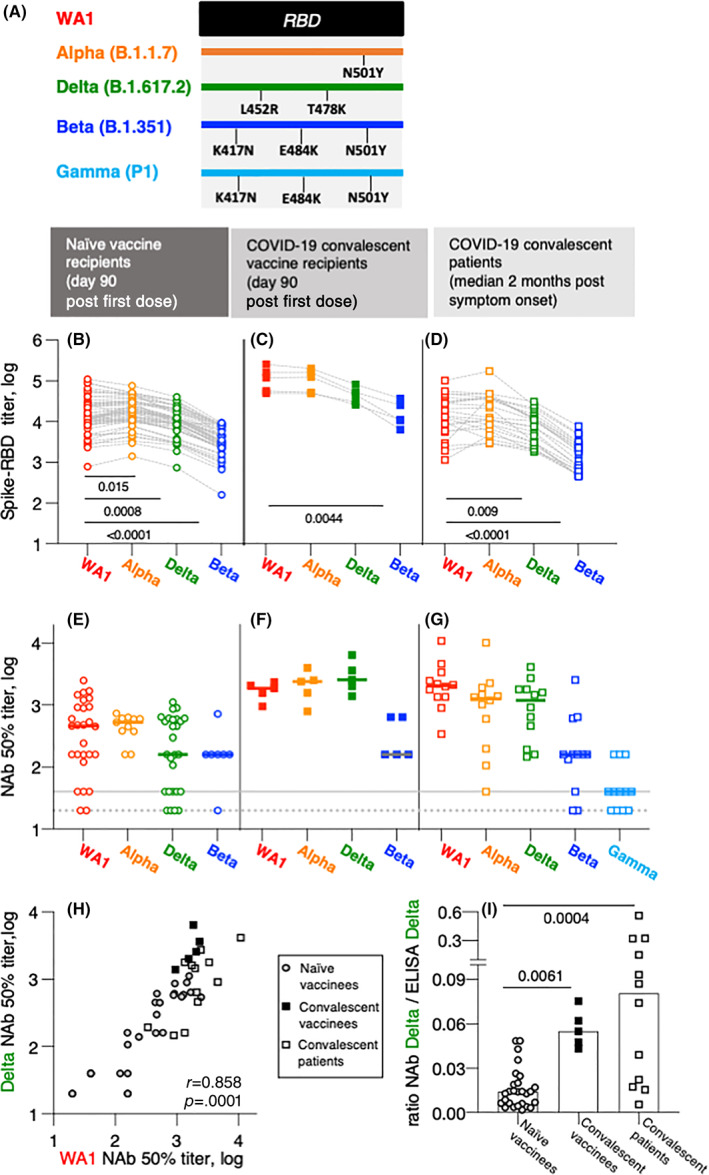
FIGURE 1.

Anti‐Spike antibody characterization in BNT162b2 mRNA vaccinated and CoV‐2 infected persons. (A) The cartoon depicts AA changes in RBD (AA 332–532). Beta and Gamma share the identical RBD but differ in several AA within Spike used in neutralization assay. (B–D) Wuhan‐Spike induced antibodies were measured by in‐house ELISA using a panel of purified Spike‐RBD proteins. (B and C) CoV‐2 naïve volunteers 1 and COVID‐19 convalescent patients 1 received the BNT162b2 mRNA vaccine and were analyzed at day 90. The convalescent vaccine recipients 1 had detectable Spike‐RBD antibodies (median titer 2.8 log, range 2.6–3.9) at the day of the first dose. (D) SARS‐CoV‐2‐infected patients 2 , 3 were analyzed at a median of 2 months postsymptom onset. Comparison between the groups were made using ANOVA Friedmann's multiple comparison test. (E–G) Neutralization was performed with the samples shown in panels (B–D) using a pseudotyped HIVNLΔEnv‐Nanoluc assay carrying a panel of Spike (AA 1–1254) proteins. Threshold of detection in gray solid line; threshold of quantification in gray dotted line. (H) Correlation of NAb to WA1 and Delta in the three cohorts described in panels (E–G). Spearman r and p value are given. (I) Ratios of Delta NAb (from panels E–G) and Delta antibody titers (panel B–D) for the different cohorts were calculated using linear values. The p values are from ANOVA Kruskal–Wallis test. ANOVA, analysis of variance
