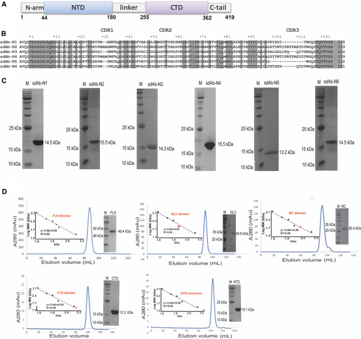
Fig. 1.

Purification of the full‐length and truncated N‐proteins, and N‐protein targeting sdAbs. (A) A schematic picture of N‐protein domain organization (B) Sequence alignment of the six nonrepetitive sdAbs. The conserved residues are highlighted and the three CDR regions are indicated by the dashed boxes. (C) 15% SDS/PAGE showing protein marker (PM) in the left lanes and purified sdAbs in the right lanes. (D) Elution profile of the full‐length and truncated N‐proteins by SEC using a Superdex 200 16/600 column (GE Healthcare, Marlborough, MA, USA). The inset shows the plotted standard curve for this column and the representative 15% SDS/PAGE showing the purified N‐proteins.
