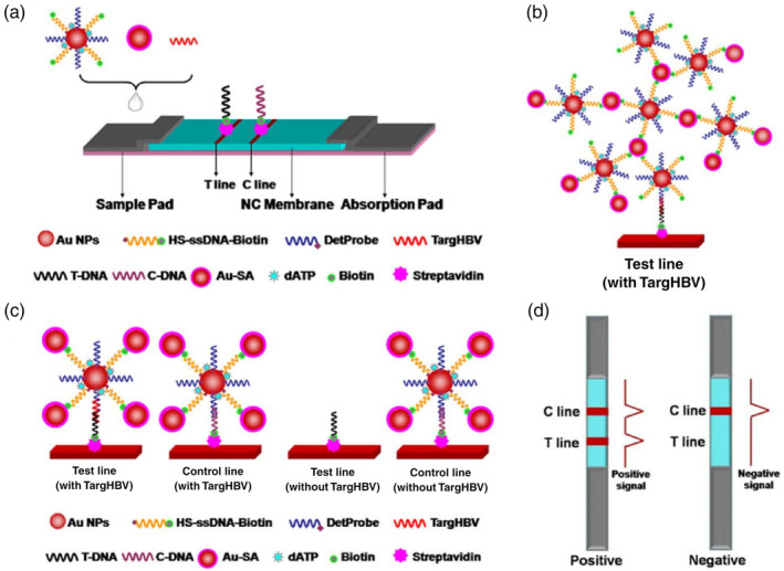
FIGURE 5.

Lateral flow test amplified by Au‐SA for nucleic acid detection. (a) Schematic representation of the configuration of the test strip. (b) Schematic illustration of the detection of nucleic acid using Au‐SA enhanced lateral flow assay. (c) Network structure on test line in the presence of targHBV. (d) Interpretation of positive and negative results. Reprinted with permission from Gao et al. (2017). Elsevier B.V
