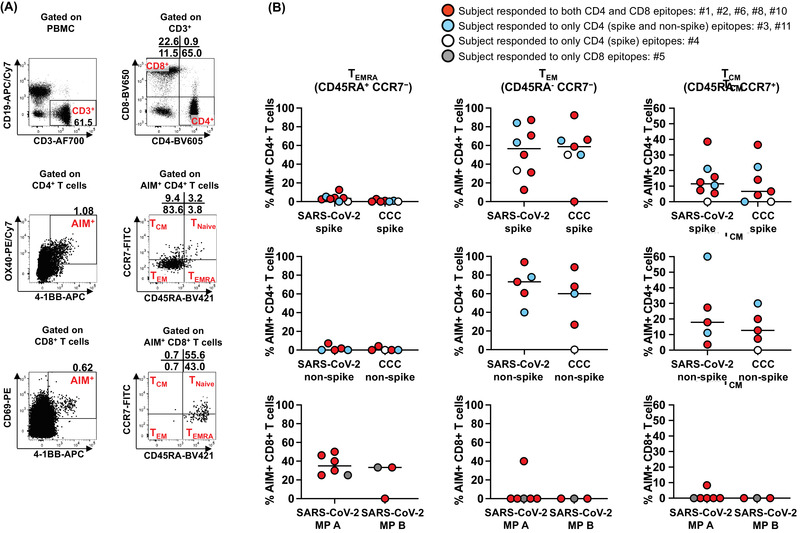
Figure 2.

Memory phenotypes of antigen‐specific, AIM+ CD4+ and CD8+ T cells in MIS‐C subjects. Memory phenotype of the antigen‐specific AIM+ CD4+ and CD8+ T cells were studied 24 h after the stimulation. (A) Gating strategies to study CD4+ and CD8+ terminally differentiated effector T cells (TEMRA), effector (TEM) and central (TCM) memory T cells. TEMRA was defined as CD45RA+ CCR7‐. Memory T cells, CD45RA‐ were further characterized by the expression of CCR7 as TEM (CD45RA‐ CCR7‐) and TCM (CD45RA‐ CCR7+). (B) Antigen‐specific CD4+ and CD8+ TEMRA, TEM, and TCM in MIS‐C subjects. Each symbol shows the percentage of TEMRA (left panels), TEM (middle panels), and TCM (right panels) in the AIM+ CD4+ or CD8+ T‐cell populations. Red circles: subjects responding to both CD4 and CD8 epitopes (#1, 2, 6, 8, and 10); blue circles: subjects responding to only CD4 (spike and nonspike) epitopes (#3 and 11); white circle: subjects responding to only CD4 (spike) epitopes (#4); gray circle: subjects responding to only CD8 epitopes (#5). Symbols represent the data derived from each individual subject. The study of 11 subjects was completed in seven independent experiments (one or two subjects/experiment depending upon enrollment). Medians were calculated and reported in the figure. Antigen‐specific CD8+ T cells showed a higher TEMRA phenotype than CD4+ T cells. In contrast, TEM and TCM were more prevalent in antigen‐specific, AIM+ CD4+ T cells than CD8+ T cells.
