FIGURE 3.

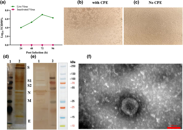
Analysis of the purified inactivated viral particles. (a) Cytopathic effect (CPE) of SARS‐CoV‐2 virus before and after inactivation. Virus titre (104–107) measured by CCID50 at three‐time points: 24, 48 and 72 h, respectively. (b) Image of Vero cell monolayer with CPE before inactivation. (c) No CPE after inactivation. (d) Samples of the purified inactivated viral particles were separated on 10% SDS‐PAGE and stained with silver nitrate: 1, molecular size markers; 2 purified viral particles. (e) Western blot: proteins on SDS‐PAGE gel were transferred onto PVDF membrane and SARS‐CoV‐2 proteins were detected using the anti‐rabbit polyclonal antibody: 1, purified viral particles and 2, molecular size markers. SDS‐PAGE and Western blot patterns show the major structural proteins: spike protein (S), nucleocapsid protein (N), membrane protein (M), and envelope protein (E). (f) Electron micrographs of negatively stained purified viral particles under TEM. Purified viral particles were negatively stained with 1% uranyl acetate and observed under TEM (TEM, EM208S, Philips, 100 kV) at 90,000 × magnification
