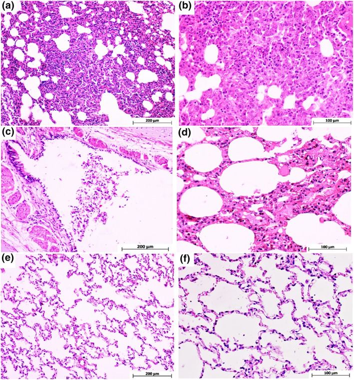
FIGURE 15.

Monkeys’ lung tissue sections stained with haematoxylin and eosin. (a and b) Different magnifications of cranial and middle lung lobes in the control group, indicating severe interstitial pneumonia with marked thickening of alveolar septa and mononuclear inflammatory cell infiltration. (c) Bronchus section from the cranial lung lobe in the animal vaccinated with 5 µg Ag + alum which shows epithelial cell necrosis and sloughing due to SARS‐Cov‐2 infection. (d) Mild interstitial inflammation of only cranial lung lobe tissue in animals vaccinated with 3 µg Ag + alum. (e and f) Show some areas of the cranial and the whole middle and caudal lung lobes in monkeys vaccinated with 3 and 5 µg Ag + alum, respectively
