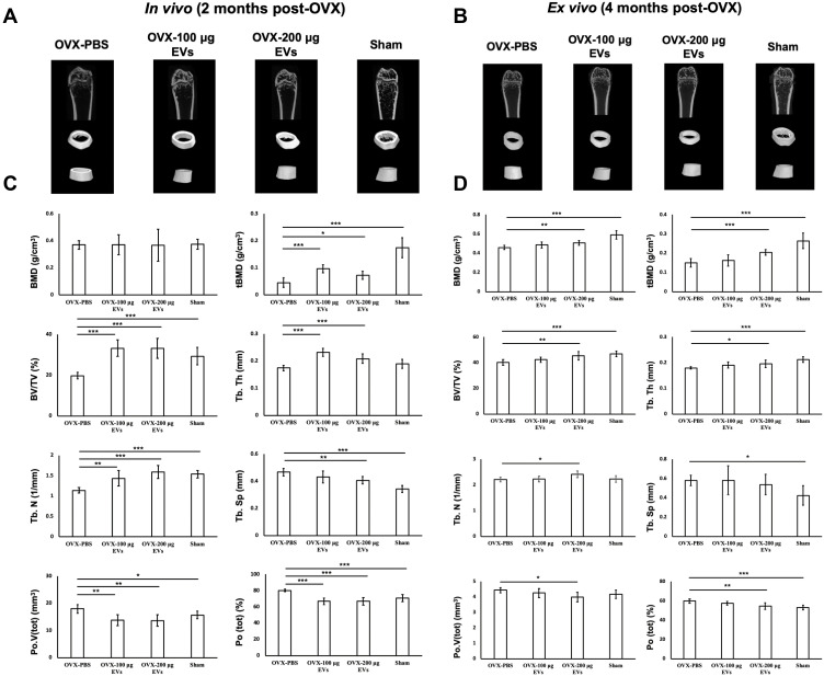
Figure 5.
In vivo and ex vivo of μCT imaging and quantitative analysis. (A and B) 3D-µCT in vivo and ex vivo images of trabecular and cortical bone microstructure in OVX-PBS, OVX-100 μg EVs, OVX-200 μg EVs, and Sham groups. (C and D) Images were further quantitatively analyzed by μCT. The parameters including BMD, tBMD, BV/TV, Tb.N, Tb.Th, Tb.Sp, Po.V(tot), and Po(tot) were acquired. Data are expressed as mean±SD (*p<0.05, **p<0.01, ***p<0.005).
Abbreviations: BMD, bone marrow density; μCT, micro-CT; OVX, ovariectomized; EV: extracellular vesicles.

