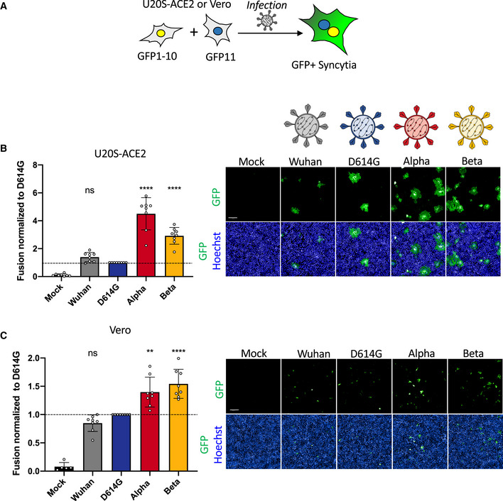
Figure 2. SARS‐CoV‐2 variant infection increases formation of syncytia in U2OS‐ACE2 and Vero GFP‐split cells.

- U2OS‐ACE2 or Vero cells expressing either GFP 1–10 or GFP 11 (1:1 ratio) were infected 24 h after plating and imaged 20 h (U2OS‐ACE2) or 48 h (Vero) post‐infection.
- Left Panel: Fusion was quantified by GFP area/ number of nuclei and normalized to D614G for U2OS‐ACE2 20 h post‐infection at MOI 0.001. Right Panel: Representative images of U2OS‐ACE2 20 h post‐infection, GFP‐Split (green), and Hoechst (blue). Top and bottom are the same images with and without Hoechst channel.
- Left Panel: Quantified fusion of Vero cells infected at MOI 0.01. Right Panel: Representative images of Vero cells 48 h post‐infection, GFP‐Split (green), and Hoechst (blue).
Data information: Scale bars: 200 µm. Data are mean ± SD of eight independent experiments. Statistical analysis: one‐way ANOVA compared with D614G reference, ns: non‐significant, **P < 0.01, ****P < 0.0001.
