
Figure 4.
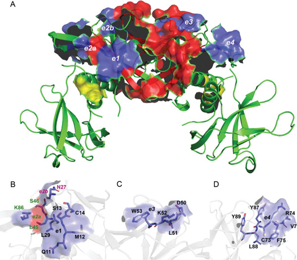
A) The SARS‐CoV‐2 Nsp9 tetramer with the interdimer and intermonomer contact surfaces highlighted in red and yellow, respectively. The blue surfaces indicate the location of the tetramer epitopes interacting with nanobodies 2NSP23 and 2NSP90. An analogous epitope pair is present on the opposite face of the tetramer. The first epitope is comprised of the surfaces e1, e2a, and e2b formed by segment [Q11‐M12‐S13‐C14] with residue L29, residue N27 and residue K86, respectively, B) and the additional contributions from L45 and S46 that are already part of the tetramer interface. The second epitope is comprised of the surfaces e3 and e4 formed by the segments C) [D50‐L51‐K52‐W53] and D) [C73‐R74‐F75‐V76 + Y87‐L88‐Y89], respectively.