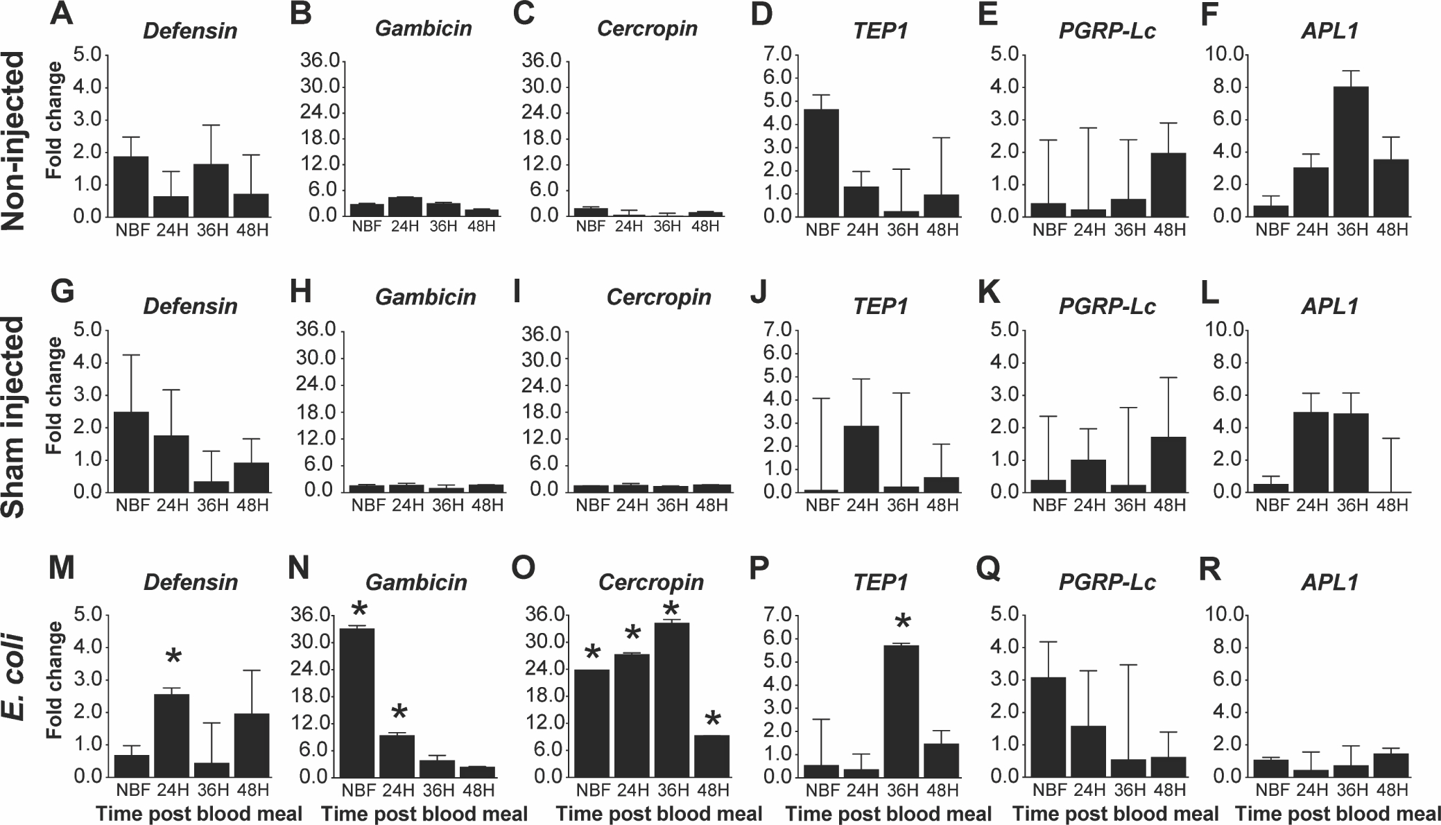
Fig. 3. Impact of increased fat body IIS on antimicrobial peptide expression following an E. coli challenge.

Graphs represent fold-change (2^(−ΔΔCt)) of TG An. stephensi transcript expression for five antimicrobial peptides (AMPs) and the thioester containing protein 1 (TEP1) relative to NTG sibling controls. Total RNA was isolated from TG and NTG An. stephensi pools (n=5 females per pool) that were either not inoculated (non-injected), inoculated with buffer only (sham injected), or inoculated with E. coli at various time points after a bloodmeal (0, 24, 36 or 48 h) to stimulate transgene expression. Transcript expression in TG mosquitoes were normalized against NTG sibling controls at each specific time points to calculate the fold change. Three biological replicates were conducted. Error bars indicate SEM and an asterisks (*) represents a significant difference (P<0.05) between the TG and NTG samples.
