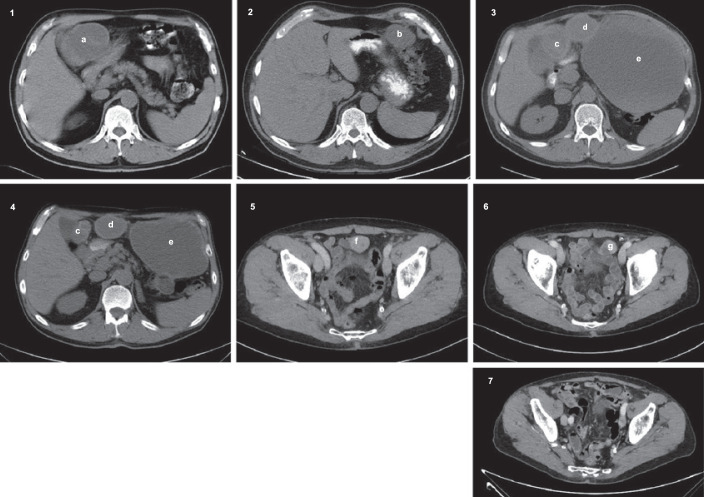
Fig. 1.
1: November 2005, primary tumour (a). 2: November 2007, image of recurrence showing 1 of 3 tumours (b). 3: August 2008, progression of disease (c–e [cystic with a peripheral solid component]). Baseline image when starting treatment with sorafenib. 4: August 2009, partial regression of tumour (c, d). The tumour (e) was not evaluated due to the cystic structure. 5: September 2017, progression of disease (f) after resection in September 2009. 6: January 2018, 15% reduction in tumour size (g) after increased dose of sorafenib. Tumour was resected in February 2018. 7. March 2021, no signs of recurrence.

