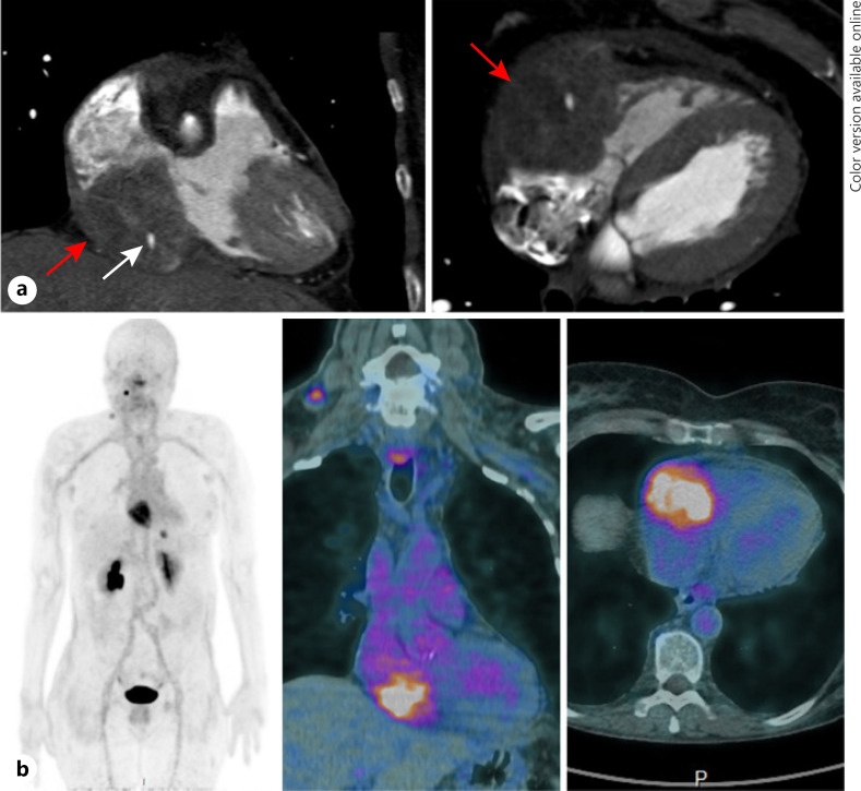
Fig. 2.
a Cardiac CT in the coronal plane (left panel) and in the sagittal plane (right panel) showing the thyroid metastasis (red arrow) encasing the right ventricle and the right atrium with the right coronary artery (white arrow) encompassed by the tumor. b Planar image (left panel) and fused images (right panel) of an immuno-PET scan using an anti-CEA bispecific antibody and a 68Ga-labeled peptide revealing cardiac uptake of the metastatic lesion with a right lateral neck and mediastinal lymphadenopathies and a liver metastasis.

