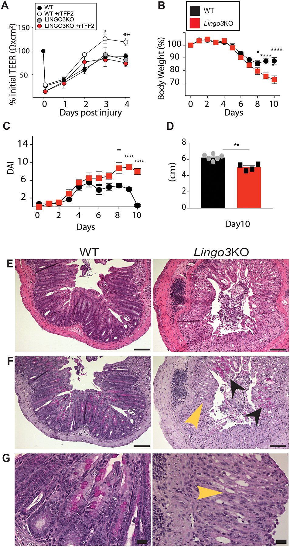
Figure 2.

LINGO3 deficiency results in poor recovery from mucosal epithelial injury. (A) Transepithelial resistance (TEER) of primary WT and LINGO3 KO nasal epithelial cells following rTFF2 (10μg/mL) or 1x PBS treatment. Following treatment of 2.5% DSS in drinking water (0–5days) and withdrawal (6–10 days), (B) Percent change in body weight (C) disease activity score (DAI) and (D) Colon lengths (cm) on day 10 of WT and LINGO3 KO mice. (E) H&E- and (F,G) PAS-stained colon sections at 10 days, 5 days following cessation of DSS (E, F: 10x, G: 40x magnification). Black arrowheads label remaining goblet cells within damaged LINGO3 LO intestinal tissue. Yellow arrowheads indicate loss of goblet cells and normal tissue architecture in KO samples. Mean ± SEM from n=4–6 samples (A) or mice/genotype (B-G). 2–3 independent experiments. *p ≤ 0.05, ** p ≤0.005, **** p ≤ 0.0001
