-
A, B
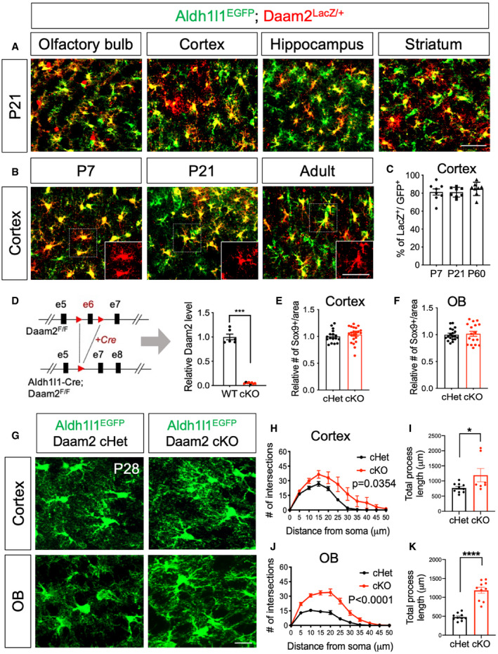
Immunostaining from Daam2LacZ/+ animals with Aldh1l1‐EGFP reporter indicates that Daam2 (LacZ+, Red) is expressed in astrocytes (Green). (A) Daam2 expression is observed in multiple brain regions at postnatal day 21. (B) Expression of Daam2 in cortex persists throughout development (P7 to adult). Scale bar: 50 μm.
-
C
Quantification of Daam2‐expressing astrocytes (LacZ+) with Aldh1l1‐EGFP‐labeled astrocytes in cortex during development. Data are presented as mean ± SEM. Two brain sections, N = 4 for each age.
-
D
Schematic of the generation of astrocyte‐specific deletion of Daam2 in Aldh1l1‐Cre and Daam2‐floxed mice. Quantitative RT–PCR in FAC‐sorted astrocytes confirms significant reduction of Daam2 transcript from Daam2 cKO mice. Experiments are performed as duplicates from N = 3–4. Data are presented as mean ± SEM. Student’s t‐test is used for statistical analysis. ***P < 0.001.
-
E, F
Quantification of astrocyte marker, Sox9+ cells in cortex (E) and OB (F) of Daam2 cHet and Daam2 cKO mice. Data are presented as mean ± SEM. 4–6 brain sections, N = 4. Student’s t‐test is used for statistical analysis.
-
G
Representative images of astrocytes labeled with Aldh1l1‐EGFP reporter in cortex and OB of Daam2 cHet and cKO mice at postnatal day 28 (P28). Scale bar: 50 μm.
-
H–K
Overall complexity of astrocytes with Aldh1l1‐EGFP reporter was measured by Sholl analysis (two‐way ANOVA) and total process length (Student’s t‐test). Data are presented as mean ± SEM. n = 6–10 cells from N = 3–5 mice per genotype, *P < 0.05, ****P < 0.0001.

