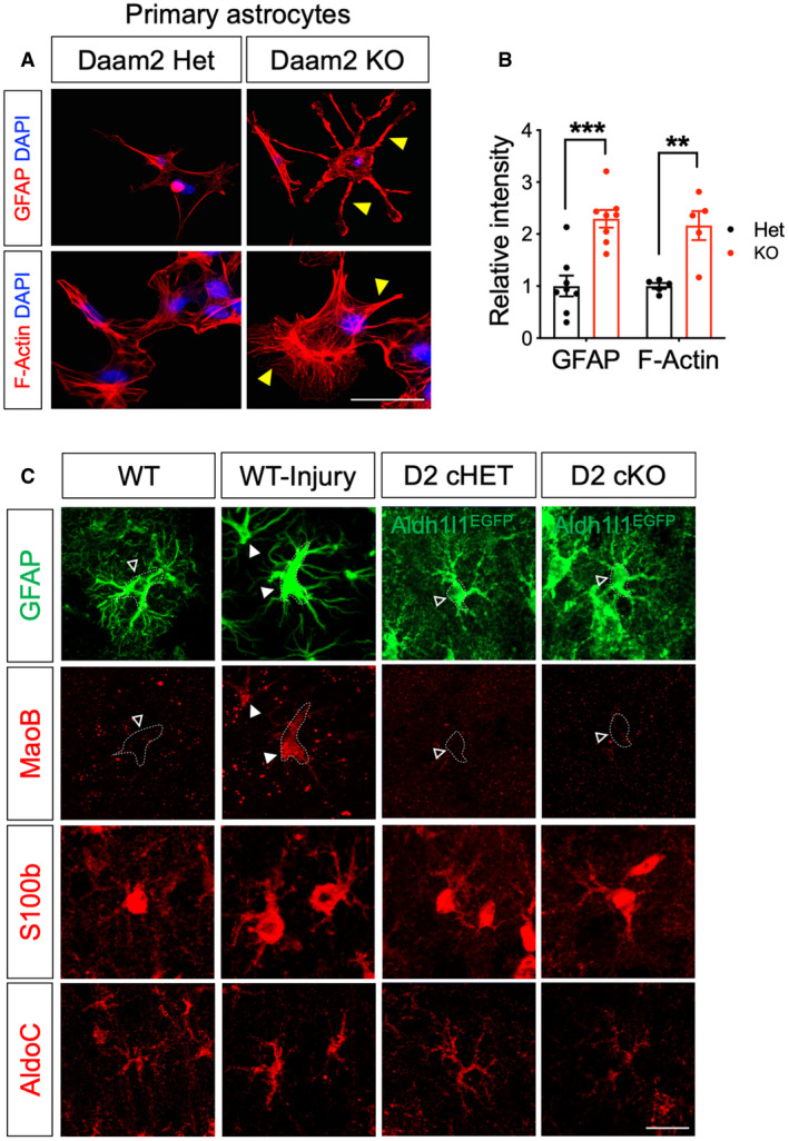
Figure EV4. Daam2‐deficient astrocytes display increased GFAP in vitro and normal level of MaoB, S100b, and AldoC expression in vivo .

-
A, BRepresentative images and quantification of immunostaining of GFAP and F‐actin in cultured primary astrocyte of Daam2 Het and KO mice. Yellow triangles indicate increased astrocytic processes in Daam2 KO astrocytes. Scale bar: 20 μm. Experiments are repeated as triplicate, and data are presented as mean ± SEM. Student’s t‐test was used, **P < 0.01, ***P < 0.001.
-
CRepresentative images of immunostaining with reactive astrocyte markers. Photothrombotic stroke was induced in the ipsilateral cortex of wild‐type mice and used as a positive control for reactive gliosis. Dotted outline indicates astrocyte cell body labeled with GFAP or Aldh1l1‐EGFP. Filled triangles and empty triangles indicate increased expression and baseline level of expression or no changes in MaoB staining, respectively. Scale bar: 20 μm.
