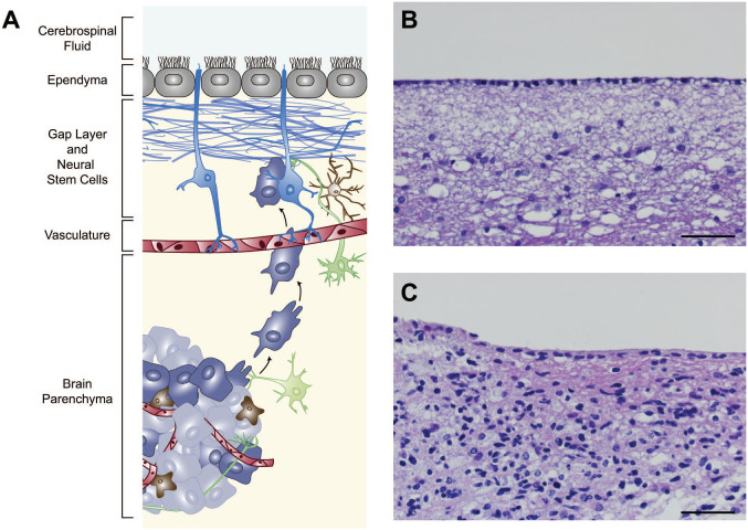
Figure 1.
The V-SVZ in schematic and in tumor histology. The mature human V-SVZ is found in the lateral walls of the lateral ventricles and is shown in a cross-sectional schematic (A) and in histological section (B). The neural stem cells that persist in this niche (shown in blue, A) have contact with the cerebrospinal fluid (at top) and the underlying vasculature (shown in red). Processes of neurons (green) and immune cells including brain-resident microglia (brown) are also found in this region, as well as the multiciliated ependymal cells (gray) which line the ventricles. The human V-SVZ is also distinguished by a gap layer (blue, bracket in A) that is largely devoid of nuclei (visible in B). Brain tumor cells (purple and gray) can be found invading this niche and migrate toward it in mouse models. In tissue from a glioblastoma patient with radiographic contact with the ventricles, abnormal cells can be seen in this region (C). Scale bars for B and C = 50 µm. Abbreviation: V-SVZ, ventricular–subventricular zone.

