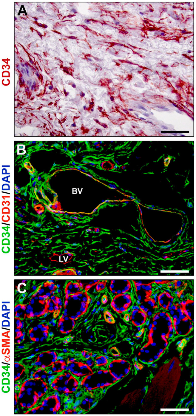
Figure 2.

Immunohistochemical localization of telocytes in human tongue connective tissue. (A) CD34 immunoperoxidase-based immunohistochemistry with hematoxylin counterstain. By means of their long and moniliform prolongations (telopodes), CD34+ telocytes form an extensive stromal cell network throughout the tongue lamina propria. (B) Double fluorescence immunohistochemistry for CD34 (green) and CD31 (red) with DAPI (blue) counterstain for nuclei. Telocytes, identifiable as CD34+CD31− stromal cells, intimately surround blood vessels showing CD34+CD31+ endothelial cells, as well as the CD34−CD31+ endothelium of initial lymphatic vessels. (C) Double fluorescence immunohistochemistry for CD34 (green) and αSMA (red) with DAPI (blue) counterstain for nuclei. CD34+αSMA− telocytes intimately encircle secretory salivary gland units outside of αSMA+ myoepithelial cells, as well as the αSMA+ pericytes of capillary vessels and smooth muscle cell layer of arterioles. Scale bar: (A–C), 50 µm. Abbreviations: αSMA, α-smooth muscle actin; BV, blood vessel; DAPI, 4′,6-diamidino-2-phenylindole; LV, lymphatic vessel.
