Figure 5.
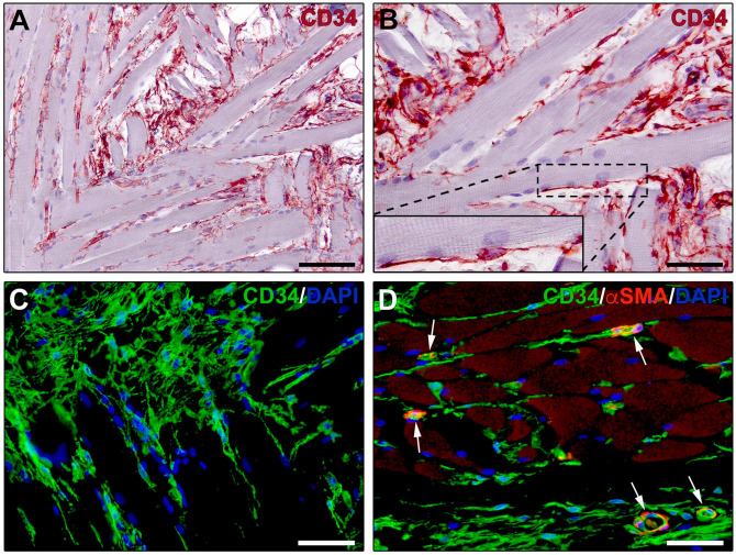
Immunohistochemical localization of telocytes in skeletal muscle interstitium. (A, B) CD34 immunoperoxidase-based immunohistochemistry with hematoxylin counterstain. (C) Fluorescence immunohistochemistry for CD34 (green) with DAPI (blue) counterstain for nuclei. CD34+ telocytes form a complex reticular network in the perimysium and endomysium. Inset in (B): higher magnification of the boxed area showing a CD34+ telocyte projecting two long and thin moniliform processes (telopodes) in close relationship with a skeletal muscle fiber. (D) Double fluorescence immunohistochemistry for CD34 (green) and αSMA (red) with DAPI (blue) counterstain. CD34+αSMA− telocytes with their telopodes surround skeletal muscle fibers and microvessels showing αSMA+ pericytes/smooth muscle cells (arrows). Scale bar: (A), 100 µm; (B–D), 50 µm. Abbreviations: αSMA, α-smooth muscle actin; DAPI, 4′,6-diamidino-2-phenylindole.

