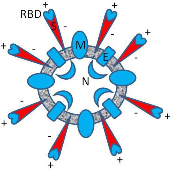
Figure 1.
Approximate diagram of the spatial distribution of electric charge in SARS-CoV-2 (B.1 variant). The resultant formal charge, estimated from the charged amino acid content of the envelope (E), the membrane (M), and the nucleocapsid protein (N) is positive (blue), respectively 2, 8, 24 elementary charge units [e]. The entire charge of spike protein (S) is negative (red), −12 [e], but locally, in the RBD, the resultant charge is positive, +7 [e], as was reported in Pawłowski’s study.5

