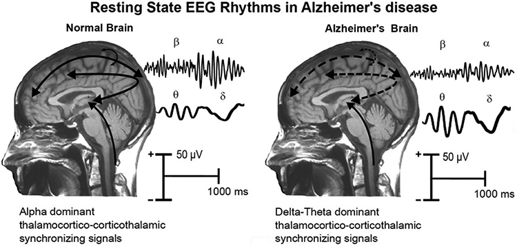
Figure 4.

Tentative physiological model of the generation of resting state eyes-closed EEG (rsEEG) rhythms in the brain of age-matched old cognitively unimpaired (CU) persons and Alzheimer’s disease (AD) patients. In the normal brain, dominant EEG rhythms are observed at alpha frequencies (8-12 Hz), which would denote the background, spontaneous synchronization around 10 Hz of neural networks regulating the fluctuation of the subject’s global arousal and consciousness states. These networks would span neural populations of cerebral cortex, thalamus, basal forebrain and brainstem, including glutamatergic, cholinergic, dopaminergic and serotoninergic parts of the reticular ascending systems. In the brain of AD patients, the amplitude of these rhythms is reduced (i.e., tonic background desynchronization) together with an amplitude increase of the pathological rsEEG slow frequencies spanning delta (< 4 Hz) and theta (4-7 Hz) rhythms. This “slowing” of rsEEG rhythms would mainly reflect a sort of thalamocortical “disconnection mode”.
