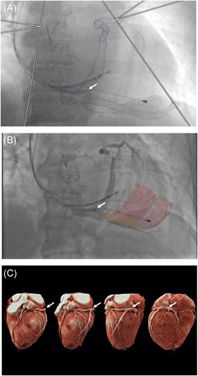
Figure 5.

CT‐derived coronary venous anatomy. (A) Overlaid onto fluoroscopy to aid operator CS cannulation. Guide catheter (arrowed) entering CS ostium. (B) Balloon venogram of corresponding coronary venous anatomy with target segments overlaid onto fluoroscopy. (C) Volume‐rendered cardiac CT angiography series delineating coronary venous anatomy (arrowed). CS, coronary sinus; CT, computed tomography
