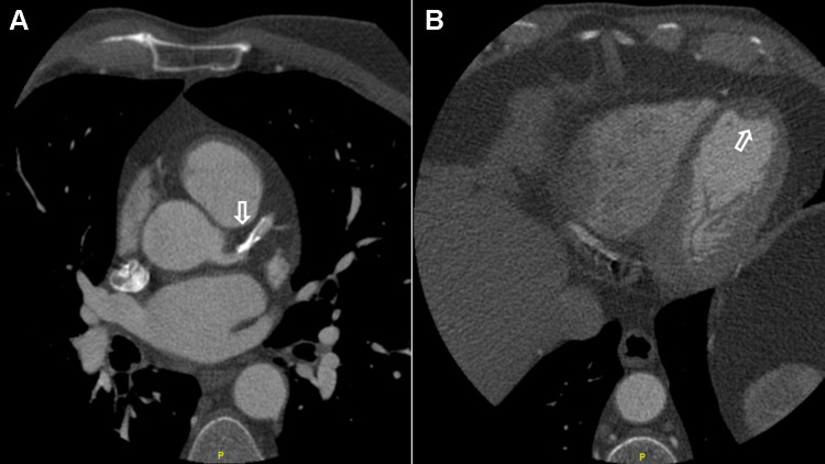
Figure 2.
The patient is 42 years old, suffering from atypical chest pain. He’s overweight, and his lab results came back positive for dyslipidemia. Cardiac CT scan performed to evaluate the presence of coronary heart disease. An ECG-gated cardiac CT acquisition was performed using an ECG-modulated radiation dose. A sublingual nitroglycerin tablet given three minutes prior to the procedure. (A) The left anterior descending artery presents a benign diffuse disease with moderate calcification and no apparent obstructive lesions (arrow). However, the presence of calcification impedes an accurate evaluation of stenosis. (B) The left ventricle is slightly dilated, the left ventricular volume at the end of diastolic is 211 mL, and the volume at the end of systole is 114 mL. The calculated ejector fraction is 46%. An organized small wall-mounted apical clot was displayed (arrow), which denotes an infarction of the old LAD territory with an organized old LV apical clot. There is an akinese of the medium and distal septum and the major part of the apex (not shown).

