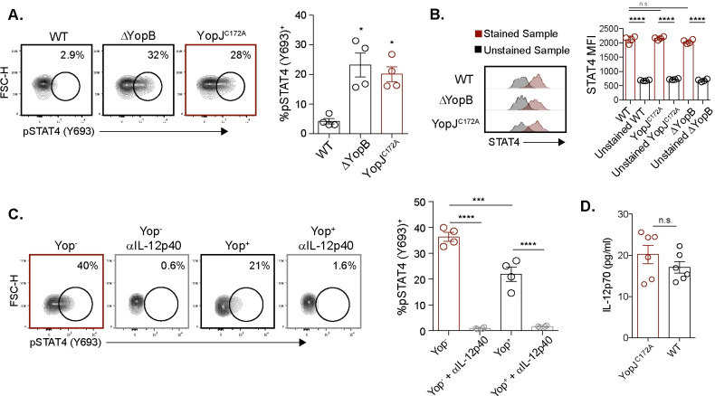
Fig 5. YopJ inhibits IL-12p40 mediated STAT4 phosphorylation.
(A) MLN cell suspensions from L. monocytogenes infected mice were stimulated with 10 MOI of WT, YopJC172A, or ΔYopB Y. pseudotuberculosis for 6 hours. Antibiotics were given 2 hours after stimulation. Vγ1.1/2- CD44hi CD27- γδ T cells were analyzed for pSTAT4 after stimulation. Representative contour plots are displayed. The graph depicts mean ± SEM and represents at least two independent experiments with 2–4 mice per group. (B) The same experimental setup was used as in (A), but Vγ1.1/2- CD44hi CD27- γδ T cells were analyzed for STAT4 protein after stimulation. Representative plots for mean fluorescent intensity (MFI) are displayed. The graph depicts mean ± SEM and represents two independent experiments with 2 mice per group. (C) MLN suspensions from L. monocytogenes infected mice were either treated or untreated with IL-12/23p40 neutralizing antibody prior to stimulation with 1 MOI of WT Yptb-βla for 6 hours. Antibiotics were given 2 hours post-stimulation. Vγ1.1/2- CD44hi CD27- γδ T cells were analyzed for pSTAT4 after stimulation. Representative contour plots are displayed. The graph depicts mean ± SEM and represents at least two independent experiments with 2–4 mice per group. (D) MLN cell suspensions from L. monocytogenes infected mice were stimulated with 10 MOI of WT or YopJC172A Y. pseudotuberculosis for 24 hours. Antibiotics were given 2 hours after stimulation. Supernatants were collected 24 hours post stimulation and IL-12p70 concentration was determined via ELISA. ****p < 0.0001, ***p < 0.001, **p < 0.01, and *p < 0.05. A repeated measures one-way ANOVA was used for (A-C). Comparisons were performed to WT Y. pseudotuberculosis in (A) and as depicted in (B-D).

