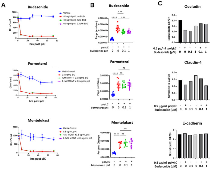
Fig 2. Budesonide, formoterol or montelukast do not impact TEER, but budesonide attenuates small molecule flux.
16HBE cells were grown to confluency (over 800 ohm) and treated with vehicle or 0.5 μg/ml polyI:C and 0.1–1μM drug. A) TEER was monitored at 6, 24, 30, 48, and 72hrs post polyI:C addition. B) At 48hrs post polyI:C, 10 μg/ml 4kDa FITC-dextran was applied apically and the amount of FITC-dextran translocation to the basal chamber was quantified 2hrs later using a fluorescent plate reader. C) 16HBE cells were treated as in A and B, but lysed in RIPA buffer for protein analysis by Western Blot. Band intensity was quantified with ImageJ and values normalized to the loading control GAPDH. Data are mean ± standard deviation. One way ANOVA followed by unpaired Tukey’s multiple comparisons test. ***p<0.01, **** p<0.0001.

