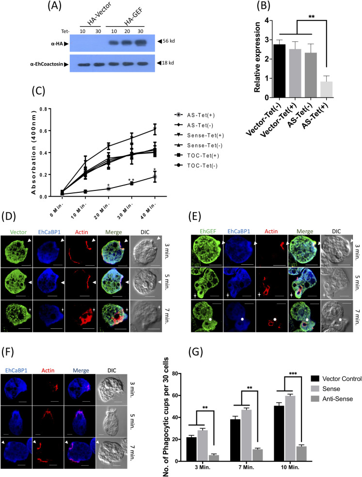
Fig 7. EhGEF regulates the phagocytosis in E. histolytica cells.
(A) HA tagged EhGEF expression was determined in presence or absence of tetracycline by immunobloting using anti HA-tag specific antibody. EhCoactosin was used as loading control. (B) EhGEF expression was quantified in anti-sense cell line of EhGEF by real-time PCR. (C) Spectrophotometric assay for erythrophagocytosis. RBC uptake assay was performed in cells expressing sense and anti-sense EhGEF constructs in presence and absence of tetracycline. RBC were incubated with indicated cell lines for different time points and the amount of RBC uptake was determined spectrophotometrically using RBC solubilisation assay as described in ‘experimental methods’. The experiments were carried out three times independently in triplicates. (D, E & F) Amoebic cells carrying vector alone (D), sense (E), and anti-sense (F) constructs in tetracycline inducible vector, were grown for 48h in presence of 30μg tetracycline and incubated with RBC for indicated times. EhGEF and EhCaBP1 were immunostained with anti HA-tag and anti EhCaBP1 antibodies followed by secondary antibodies conjugated with Alexa-488 and Alexa-405 respectively. EhActin was visualized by TRITC-phalloidin staining. Solid arrow showed the phagocytic cups. (G) Quantitative analysis of phagocytic cups. Randomly 30 cells were selected in three sets for each experiment and number of phagocytic cups present in selected cells were counted for indicated cell lines.Scale bar indicated 10μm, DIC is differential interference contrast. ANOVA test was used for statistical comparisons.*p-value≤0.05, **p-value≤0.005, ***p-value≤0.0005.

