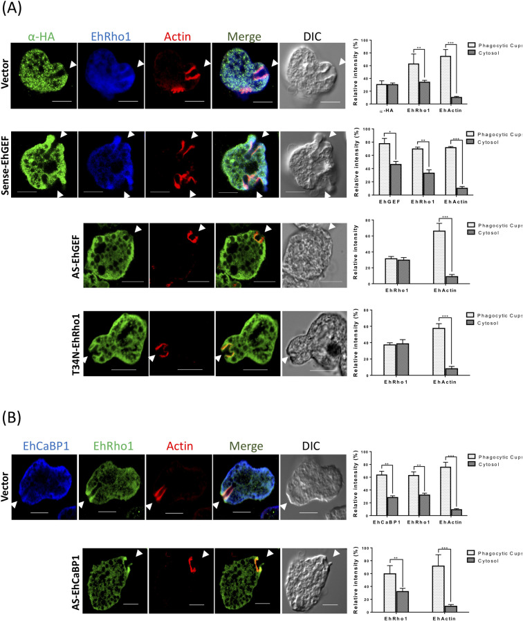
Fig 8. EhRho1 recruited through EhGEF.
(A) E. histolytica cells expressing indicated constructs were grown in presence of 30μg/ml tetracycline and incubated with RBC for 5 min at 37°C. Cells were fixed and immunostained with indicated protein-specific antibodies. Alexa-488 (green) and pacific blue-410 (blue) were used for immunostaining of proteins and F-actin was stained with TRITC phalloidin (red). Arrowheads indicated phagocytic cups with enrichment of indicated proteins and asterisk marks the just closed cup before scission and star represent newly form phagosomes. Transfectants downregulated for EhGEF expression (EhGEF-AS) and overexpressing T34N mutant of EhRho1, both showed dominant negative phenotype. Graphs represent the quantitative analysis of fluorescent signal from cytosol and phagocytic cups of immunostained images of EhGEF, EhRho1 and EhActin containing cells. Five random regions were selected in five cells for analysis of fluorescent signal from cytosol and phagocytic cups of indicated cells and average fluorescent intensity was calculated for each region (N = 5, bar represent standard error). Relative intensity was calculated by assuming intensity as 100% for each marker separately. (B) Immunofluorescence images of E. histolytica cells containing indicated constructs during erythrophagocytosis. Cells were stained with anti-EhRho1 or anti-CaBP1 antibodies followed by Alexa-405 or Alexa-488 secondary antibodies. Actin was stain with TRITC-phalloidin. The recruitment of EhRho1 is independent of EhCaBP1 mediated signalling pathway. ANOVA test was used for statistical comparisons.*p-value≤0.05, **p-value≤0.005, ***p-value≤0.0005.Bar represent 10μm, DIC is differential interference contrast.

