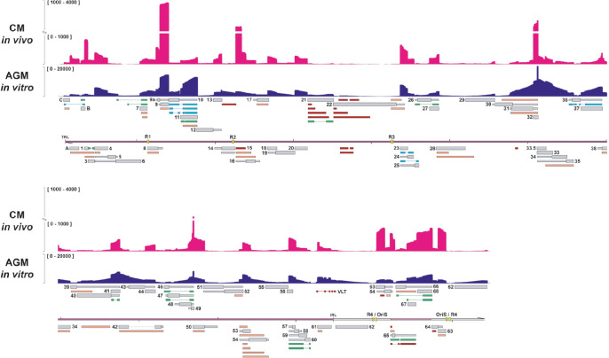
Fig 5. Comparison of the lytic SVV transcriptome in vitro and in vivo.
PCR-cDNA sequencing of RNA obtained from lung tissue of cynomolgous macaques at 3 days post-infection with SVV strain Delta reveals the lytic transcriptome in vivo. Coverage plots showing PCR-cDNA-seq of SVV-infected CM lung tissue in vivo (magenta) and AGM BS-C-1 cells in vitro (dark blue), with Y-axis indicating the unstranded read depth. Canonical (grey), variant (orange), splice variant (green), fusion transcripts (blue) and non-coding (red) RNAs are indicated. Double black lines represent the SVV genome, with UL in purple fill, US in pink and IRS/TRS no fill, ticks indicate 10 kb intervals and the reiterative repeat regions R1 to R4 and both copies of OriS are indicated as yellow boxes on the genome track.

