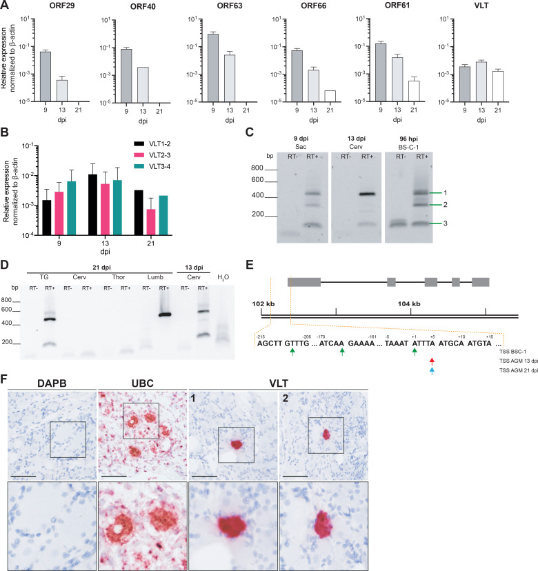
Fig 8. Characterization of VLT expression in ganglia during establishment of latency.
(A-B) Detection of lytic SVV RNAs and VLT by qPCR on cDNA obtained from pooled DRG (n = 5 anatomical levels) of SVV-infected AGMs at 9 (n = 2 animals), 13 (n = 2 animals) and 21 (n = 1 animal) days post-infection. Data represent mean ± SEM. (B) Comparison of three VLT exon-spanning primer-probe pairs. (C) RT-PCR showing VLT expression during lytic infection in cell culture (BS-C-1) at 96 hpi, acute sacral (Sac) DRG infection (9 dpi) and transition to latency in cervical ganglia (13 dpi) using primers located in SVV VLT exon 1 and exon 4. Band numbers correspond to isoforms depicted in Fig 7B. (D) Nested RT-PCR on trigeminal ganglia (TG) and DRG (Cerv = cervical, Thor = thoracic, Lumb = lumbar) obtained from SVV-infected AGM at 13 and 21 dpi, respectively. (E) Transcription start sites of VLT in BS-C-1 cells (top row) or AGM ganglia at 13 dpi (middle row) and 21 dpi (bottom row), as determined by 5’RACE. (F) Detection of VLT RNA (red signal) by in situ hybridization in DRG neurons of a SVV-infected rhesus macaques at 21 dpi. Probes directed to bacterial gene DAPB and abundantly expressed ubiquitin C (UBC) were included as negative and positive controls, respectively, and sections were counterstained with hematoxylin. Representative images are shown for RM 2207 (lumbar; DAPB, UBC and VLT image 1) and RM 9021 sacral (VLT image 2). Scale bar indicates 50 μm, magnification 40x (upper), inset: 2.5x digital zoom (lower).

