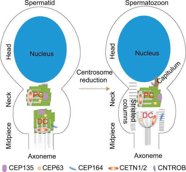
Figure 1.

The sperm centrosome is remodeled during spermatogenesis. Initially, there are two centrioles located in the neck of spermatid cell. Both the PC, which articulates with the sperm nucleus, and the DC are composed of barrel-shaped microtubules. Later during spermatogenesis, the DC is remodeled through centrosome reduction into a structure consisting of splayed microtubules, and the PCM transforms into the capitulum and striated columns. The centriolar lumen protein CETN1/2 and the PCM protein CEP63 localize at the DC, while the centriole wall protein CEP135 and the appendage protein CEP164 are lost. The protein CEP164 instead localizes to the striated columns. The PC is slightly altered in mature spermatozoa. It maintains the typical centriole structure and the centriolar proteins CEP135 and CETN1/2, the PCM protein CEP63, and the appendage protein CEP164. The centriole wall protein CNTROB is missed from the centriole wall but localizes to the capitulum.
