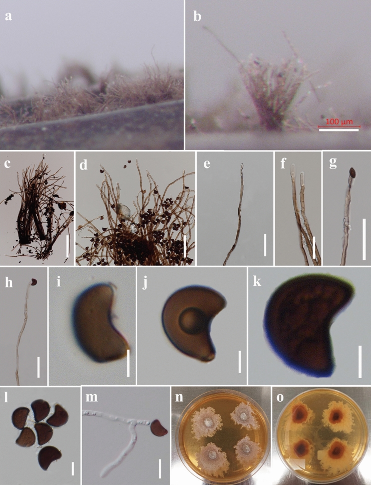
Fig. 157.
Melanographium smilacis (MFLU 21-0075, holotype). a, b Appearance of colonies on natural substrate. c, d Squash mount of conidiophores with attached conidia. e, f Close-up of conidiophores showing septation with paler at the apex. g, h Apex of conidiophores with developing conidia. i–l Conidia with a basal scar. m Germinated conidium. n, o Culture on MEA from surface and reverse. Scale bars: b = 100 µm, c, d = 200 µm, e–h = 50 µm, i–l = 5 µm, m = 10 µm

