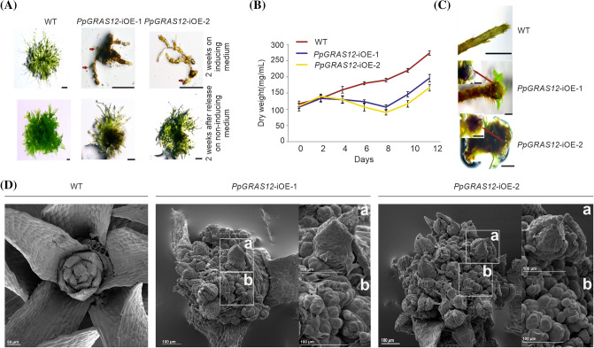
Fig. 3.
Phenotypic analysis of the PpGRAS12-iOE lines. a Equal amounts of protonema tissues from the WT and both PpGRAS12-iOE lines were spotted on standard solid growth medium supplemented with 2 µM ß-estradiol. Upper panel: protonema tissue after growth for 14 days on the medium supplemented with 2 µM ß-estradiol. Lower panel: 14 days after growth on inducing medium protonema tissue was transferred onto standard growth medium without inducer for 2 weeks. Red arrows indicate viable green cells. Scale bars: 1 mm. b PpGRAS12-iOE lines and WT were grown in standard liquid medium. Protonema from the PpGRAS12-iOE lines and WT were induced with 2 µM of ß-estradiol and dry weight of samples was measured every 2 days for a period of 12 days. Error bars indicate mean values ± SE (n = 3). c Formation of abnormal structures at the tip of both PpGRAS12-iOE lines. Scale bar: 1 mm for the WT and 0.5 mm for the mutants. d SEM analysis of PpGRAS12-iOE lines. The formation of supernumerary apical meristems in the PpGRAS12-iOE lines upon the induction with 2 µM of ß-estradiol. Box a: a leafy gametophore that was formed from an individual apical meristem. Box b: enlarged supernumerary apical meristems

