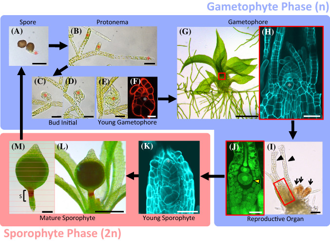
Fig. 2.
Life cycle and primary stem cells in P. patens. a, b Asterisks indicate protonemal apical cells. Spore germination (a) and growing protonemata (b). c–h Asterisks indicate shoot apical cells. Growing bud initial (c–e), and optical section (single plane image from confocal microscope) of bud initial almost at the same stage as e (f). Growing gametophore (g) and optical section of the red square inset in panel g (h). i, j The sexual organ at the top of the gametophore. Black arrowheads show archegonia, and black arrows show antheridia (I). The basal part of the archegonia indicated by the red square inset in i (j). The yellow arrowhead indicates an egg cell. k developing embryo. The asterisk shows the sporophytic apical cell. l, m Mature sporophyte. ‘S’ indicates seta region (m). Scale bars 50 μm (a–e, h, i, k), 20 μm (f, j), 500 μm (g, l), 200 μm (m)

