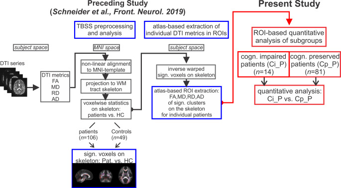
Fig. 1.
Study design: built on atlas-based region of interest (ROI) extraction the present study investigated (red framed elements) quantitative diffusion tensor imaging (DTI) metrics of cognitively impaired patients (Ci_P, n = 14) in comparison to cognitively preserved patients (Cp_P, n = 81). It was based on the results of a preceding tract-based spatial statistics (TBSS) analysis (blue framed elements) which yielded individual fractional anisotropy (FA), mean diffusivity (MD), radial diffusivity (RD) and axial diffusivity (AD) values in clusters on the white matter (WM) skeleton in which significant differences between patients and healthy controls (HC) have been detected [15]. MNI Montreal Neurological Institute, cogn. cognitive, Pat. patients, sign. significant, vs. versus (compared against)

