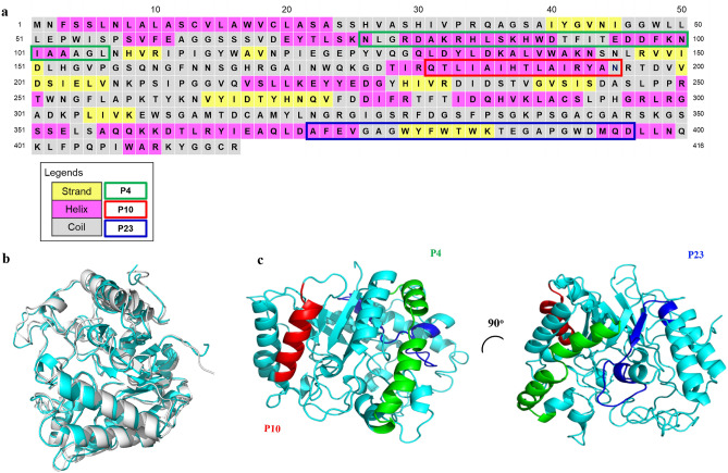
Figure 1.
Primary sequence and predicted secondary structure elements of gp43 from Paracoccidioides brasiliensis strain B339 highlighting the three peptides studied here (P4, P10 and P23). (a) The primary structure was obtained at GenBank access number AAG36697.1. Secondary structure elements of gp43 were predicted by PSIPRED and are indicated by pink, yellow and grey cells where color refers to, respectively, α-helix, β-strand and random-coil structures. Green, red and blue outlines represent P4, P10 and P23 primary sequences, respectively. (b) The structural model of gp43 was generated by using AlphaFold software (cyan), which was aligned with the β-glucanase structure (gray). (c) Positions of P4, P10 and P23 in the structural model of gp43 are depicted in green, red and blue, respectively. Superposition of both structures in B was generated by PyMOL (The PyMOL Molecular Graphics System, Version 1.2r3pre, Schrödinger, LLC.).

