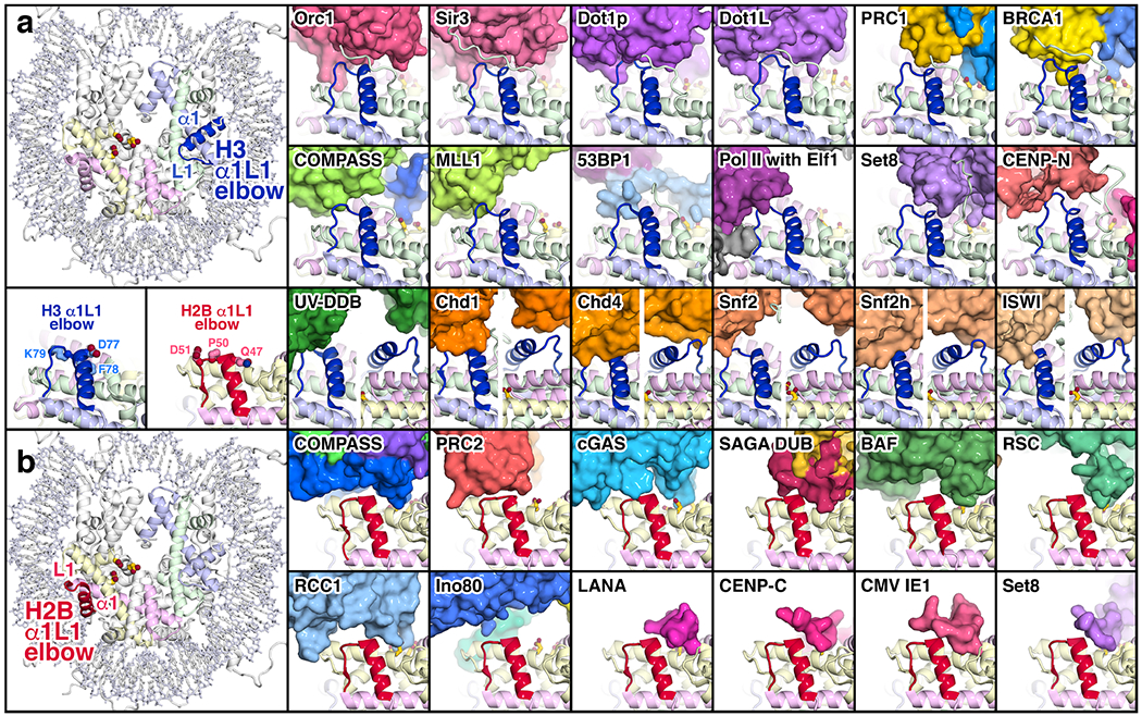
Fig. 3:

Interactions of chromatin proteins with the histone H3 and H2B α1L1 elbows. (a) Interactions with the H3 α1L1 elbow. The location of the H3 α1L1 elbow is shown on the left in the nucleosome crystal structure (PDB id 1KX5 [70]) together with the H2A acidic patch residues E61, D90 and E92. To the right are shown proteins and protein complexes in surface representation that interact with the H3 α1L1 elbow (blue): Orc1 (PDB id 6OM3 [31]), Sir3 (PDB id 3TU4 [6]), Dot1p (PDB id 7K6Q [32]), Dot1L (PDB id 6NQA [21]), PRC1 (PDB id 4R8P [5]), BRCA1 (PDB id 7JZV [30]), COMPASS (PDB id 6VEN [27]), MLL1 (PDB id 6KIU [36]), 53BP1 (PDB id 5KGF [69]), PolII elongation complex with Elf1 (PDB id 6IR9 [52]), Set8 (PDB id 7D1Z [29]), CENP-N (PDB id 6MUP [71]), UV-DDB (PDB id 6R8Y [62]), Chd1 (PDB id 6FTX [38]), Chd4 (PDB id 6RYR [39]), Snf2 (PDB id 5X0Y [40]), Snf2h(PDB id 6NE3 [41]) and ISWI (PDB id 6K1P [42]). In the CENP-CN structure, the α1L1 elbow of the centromeric histone variant, CENP-A, contains two extra residues compared to the histone H3 it replaces. The histones are shown in cartoon representation, and the nucleosomal DNA omitted for clarity. The H3 α1L1 elbow as it occurs in the nucleosome crystal structure is shown in blue in the same orientation together with elbow residues D77, F78 and K79 on the left below the full nucleosome structure. An additional view from the back is provided for Chd1, Chd4, Snf2, Snf2h and ISWI. (b) Interactions with the H2B α1L1 elbow. The location of the H2B α1L1 elbow is shown on the left in the nucleosome crystal structure (PDB id 1k5x) together with the H2A acidic patch residues E61, D90 and E92. Proteins and protein complexes shown that interact with the H2B α1L1 elbow (red) are COMPASS (PDB id 6VEN [27]), PRC2 (PDB id 6WKR [45]), cGAS (PDB id 7JO9 [14]), SAGA DUB (PDB id 4ZUX [44]), BAF (PDB id 6LTJ [24]), RSC (PDB id 6TDA [25]), RCC1 (PDB id 3MVD [3]), Ino80 (PDB id 6FML [46]), LANA (PDB id 1ZLA [2]), CENP-C (PDB id 4X23 [4]), CMV IE1 (PDB id 5E5A [43]) and Set8 (PDB id 7D1Z [29]). The H2B α1L1 elbow as it occurs in the nucleosome crystal structure is shown in red in the same orientation together with elbow residues Q47, P50 and D51 on the left above the full nucleosome structure.
