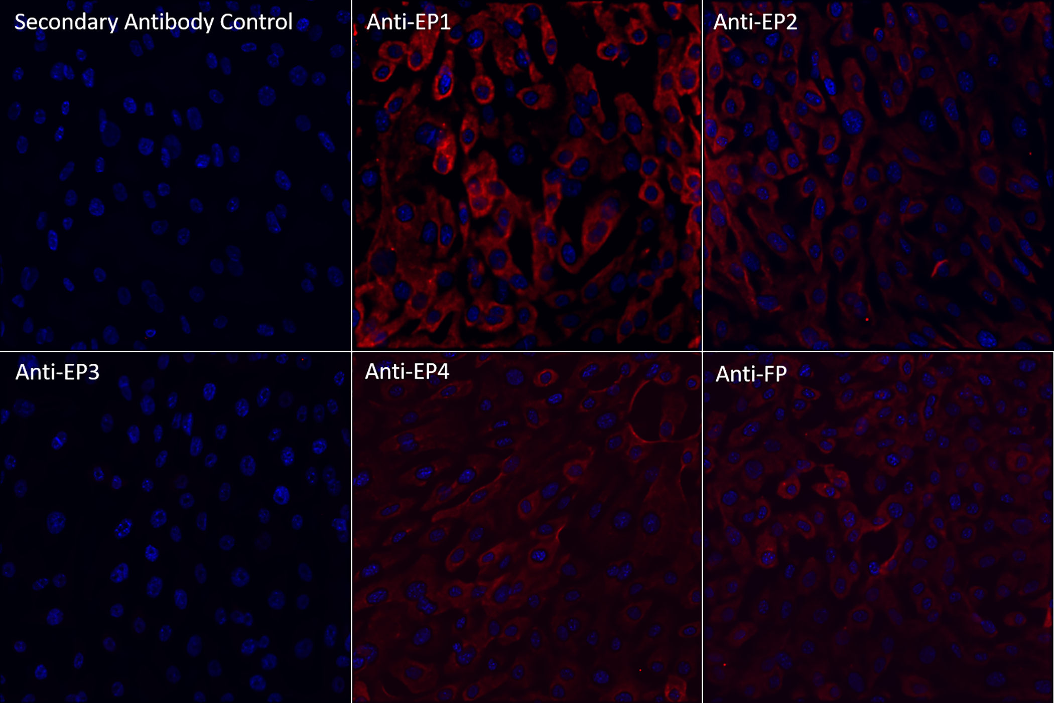
Figure 1:

Fluorescent microscopy images (40x) of HMGECs stained with primary antibodies against PGE2 (EP1, EP2, EP3, or EP4) or PGF2α (FP) receptors and counterstained with DAPI (blue) after 24 hours of culture in differentiation media containing DMEM/F12, 10 ng/ml EGF, 2% FBS, and 50 μM rosiglitazone. Positive signal for each primary antibody (pseudocolored red) was detected for EP1, EP2, EP4, and FP receptors.
PGE2: prostaglandin E2
PGF2α: prostaglandin F2α
