
Figure 2:
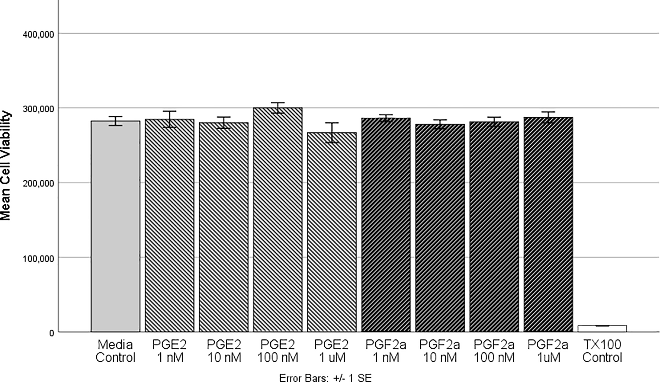
HMGECs were differentiated for two days (see Methods) prior to exposure to PGE2 or PGF2α for 3 hours. After incubation, cell viability was assessed with a luminescent ATP-based assay. There were no significant differences between the vehicle control and any of the PGE2 or PGF2α concentrations, suggesting that these prostaglandins at physiologic concentrations do not alter cell viability. Further there were no differences between PGE2 or PGF2α. Triton-X 100 (1%) was used as a positive control, which differed significantly from all other concentrations (p < 0.001). n = 6 per condition
HMGEC: human meibomian gland epithelial cell
PGE2: prostaglandin E2
PGF2α: prostaglandin F2α